|
A Happy Dog is a Healthy Dog so worm and flea regularly according to your veterinary's instructions.
|
Worming and Parasites
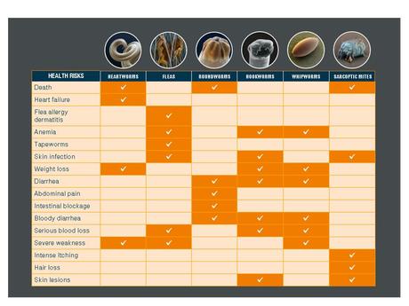
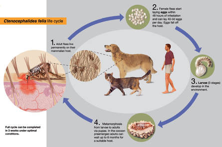
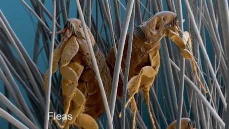
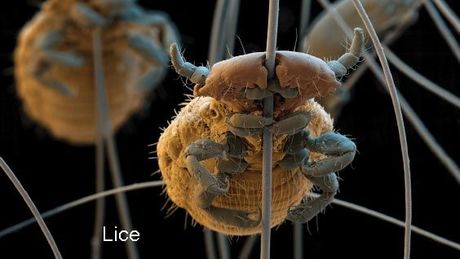
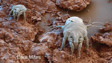
Worming All dogs will have worms at some point in their lives, with puppies being most at risk. An untreated worm infestation can lead to a loss of condition in the adult dog and quite serious illness in puppies, as well as putting human health at risk (however small this risk may be). As a responsible dog owner, it is important for you to worm your dog regularly. Dogs with worms may not show signs of illness, except when the worms are present in large numbers. Puppies are most at risk from worm infections. Worms are passed from the mother before birth, and after through the milk. Infestation may cause weight loss, vomiting, diarrhoea, a swollen abdomen and, in extreme circumstances, death. Puppies should be wormed from two weeks of age at two weekly intervals until they are twelve weeks of age, then every month until they are six months of age. Worming should continue at least three times a year with a recommended veterinary preparation for the rest of the dog’s life. Pregnant bitches should be wormed at the time of mating and again when the puppies are one week old. Most worms will live in the intestine and feed on partly digested food. ECTOPARASITES External parasites or ectoparasites naturally are the better known group of parasites as the symptoms are so obvious. Infestation happens through direct transmission between animals or through eggs that have been shed by an animal. Typical symptoms are itching, scratching, hair loss, ulcerated skin and others depending on the species. FLEAS One flea can bite as much as 400 times a day and skin irritation due to its saliva can cause significant suffering for the animal. Another, and even more serious threat for the animal, is transmission of pathogens like Bartonella or tapeworm both of which can also infect humans. The fact that it takes a lot of time and diligence to get rid of fleas once they are in the house makes prevention even more important. LICE Lice on dogs are a widespread health problem. The canine chewing louse Trichodectes canis is found worldwide and is known to transmit the dog tapeworm. If a dog is properly protected and healthy lice infestation rarely occurs. MITES Parasitic mites are usually very small eight-legged organisms which may cause skin diseases. MITE SARCOPTES SCABIEI/ NOTOEDRES CATI Sarcoptes and Notoedres mites are very similar regarding life cycle and disease symptoms but they affect different hosts:
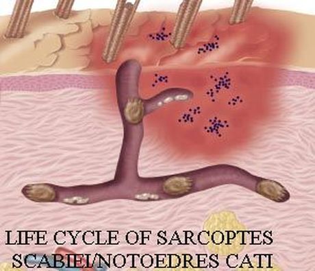
For both species transmission to humans is possible. LIFE CYCLE OF SARCOPTES SCABIEI/NOTOEDRES CATI Female mites burrow into skin and lay eggs several times. The larvae hatch from eggs and molt to nymphs which change into the adult form inside the burrow. The entire life cycle takes 2-3 weeks. Symptoms of mange in dogs and in cats:
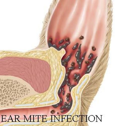
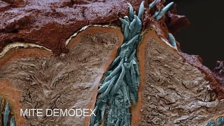
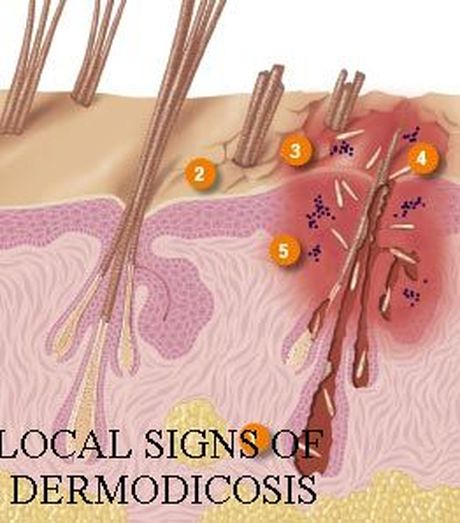
ENDOPARASITES Worms in dogs and cats live in the inner organs or bloodstream of the animal and are for obvious reasons more difficult to diagnose than ectoparasites. They can affect heart, lung, skin or the gastro-intestinal system and with some parasites infestation can be lethal if not treated in time. EAR MITE OTODECTES Otodectes cynotis, also known as ear mite, is the most common mite found in cats and dogs and is usually transmitted via direct contact with an infected animal, from dog to dog and from cat to cat. EAR MITE INFECTION It is involved in 50% of external ear infections in dogs and cats1and very common in young animals. Typically it causes an infection of the outer ear canal which often leads to inflammation or even allergic reactions. MITE DEMODEX Demodex canis are microscopic, lancet-shaped mites living in hair follicles. In low numbers, Demodex mites are part of the normal skin fauna and nursing puppies acquire D. canis from their mother in the first few days of life. If a dog’s immune system is impaired it can lead to canine demodicosis, a challenging skin disease. Local signs can vary widely, but include multifocal alopecia
Classification of demodicosis based on the symptoms of the disease:
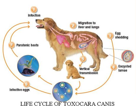
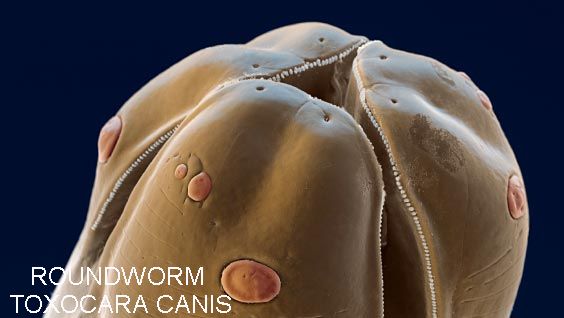
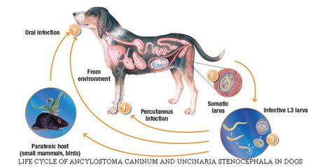
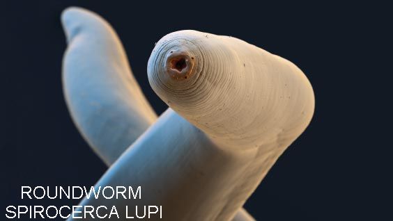
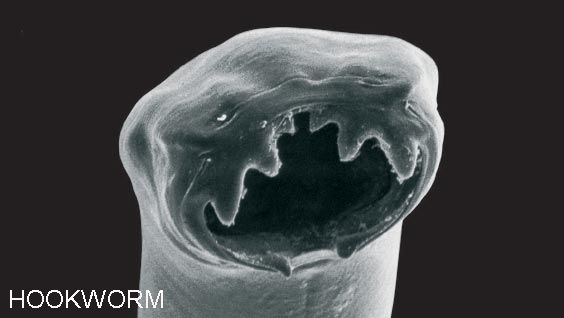
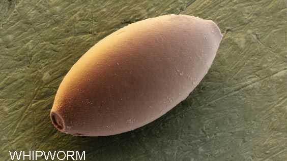
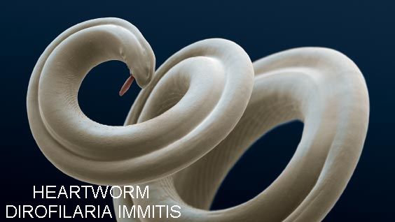
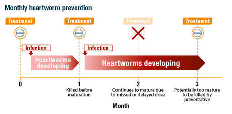
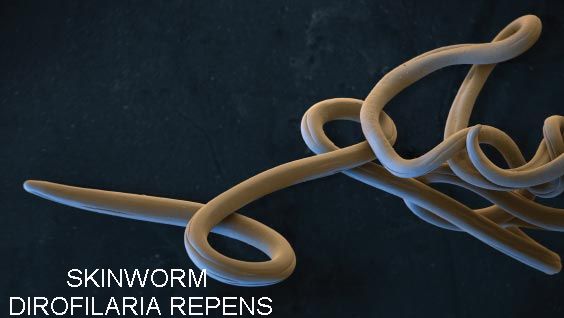
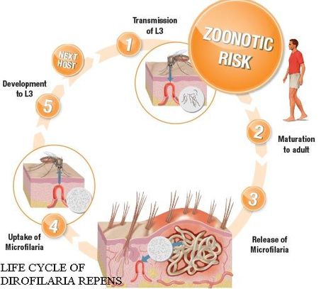
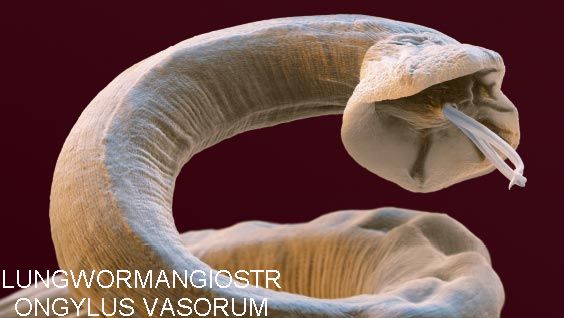
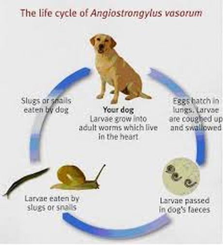
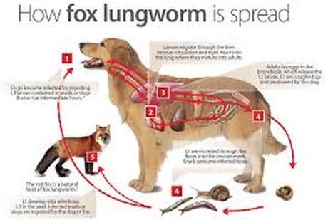
INTESTINAL WORMS Intestinal worms are classified into 4 types: tapeworms and the nematodes roundworms, hookworms and whipworms. Between the 4 types there are differences regarding intermediate hosts, route of infection and zoonotic risk for humans. ORAL DEWORMING AGAINST INTESTINAL WORMS Quarterly deworming with oral products kills worms through a short dosing. However, infection may occur immediately after treatment and there is enough time for larvae to develop and cause damage to the pet’s health until the next treatment. Infection may occur immediately after deworming and there is enough time for larvae to develop and cause damage to the pet’s health until the next treatment. ROUNDWORM TOXOCARA CANIS Toxocara canis, also known as dog roundworm, is found worldwide. Development happens via excreted eggs in which an infective larva develops. Upon oral uptake this develops via further molds to adult stages. The developing stages L4 and immature adult in the host are susceptible to some treatments. LIFE CYCLE OF TOXOCARA CANIS Dogs become infected by ingesting infective eggs from the soil or vegetation, or infective larvae in small mammals or birds. Ingested eggs hatch in the dog‘s gut. Larvae penetrate the bowel wall and migrate to travel to lungs. From the lungs, larvae will either undergo tracheal or somatic migration. Eggs are shed in the faeces, contaminating the environment. Some larvae encyst in body tissues, become dormant, and can be reactivated during pregnancy and migrate to developing pups in utero or via the mammary glands of the mother. Larvae can be transferred to puppies before birth or through nursing. T. canis eggs become infective in 2 to 7 weeks depending on environmental conditions and remain infective in the soil for years. T. canis eggs are shed through faeces and can stay infective in soil for years, constituting a zoonotic risk especially for elderly and children, e.g. in playgrounds. If a human is infected larvae may reach body tissues such as the brain or the eye, a serious disease called Visceral or Ocular Larvae Migrans. Regular, ideally monthly worming is crucial to control worms before reaching the adult stage. If worms mature they produce eggs, which are shed into the environment through the dog’s faeces and create a risk of re-infestation. ROUNDWORM SPIROCERCA LUPI Spirocerca lupi is a nematode, which is mainly affecting dogs. Intermediate host is a dung beetle, reservoir hosts are wildlife canids, such as foxes and wolves. As it occurs mainly in warm climates it is a risk to be concerned for dogs travelling to southern European countries. Spirocerca lupi affects the oesophagus and aorta of the dog. Early symptoms are often very subtle (regurgitation, vomiting, coughing, dyspnoea) but aortic nodules may lead to death by aneurism rupture. Oesophageal nodules may also become cancerous. HOOKWORMS The hookworm resides in the small intestine of its host, which can be a dog, cat or human. Infections may occur by various routes: percutaneous or oral. Typical signs and symptoms of a hookworm infection are inflammation in the small intestine, anaemia or skin irritation. When hookworm larvae penetrate human skin an itching disease called cutaneous larva migrans may occur which shows as wormlike burrows underneath the skin (“creeping eruption”). Typical hookworms in dogs are Ancylostoma caninum andUncinaria stenocephala, typical in cats is Ancylostoma tubaeforme. All three are very similar regarding transmission, paratenic hosts, symptoms and zoonotic risk. LIFE CYCLE OF ANCYLOSTOMA CANINUM AND UNCINARIA STENOCEPHALA IN DOGS Dogs become infected by ingesting infective larvae from the soil, by eating paratenic hosts or by hookworm larvae entering their host by active penetration of the skin, especially between the footpads. Eggs are shed in the faeces and hatch into infectious larvae in the environment. Larvae can also migrate during pregnancy to developing pups in utero or via the mammary glands of the mother. WHIPWORMS The whipworm Trichuris vulpis, a blood feeding nematode, is a significant health risk, especially for younger dogs, and rarely found in cats. It resides in the beginning of the large intestine (caecum) with the anterior end of the body embedded in the mucosa. In young dogs infections may cause reduced growth and wasting. Massive infection can result in mucus and blood in the faeces, foul smel and/or concurrent anaemia. For effective control monthly treatment is important. HEARTWORMS Regarding heartworms in dogs, monthly prevention is very important because once the animal is infected only infective larvae and developing stages can be killed to avoid maturation into adult forms. Treatment of matured worms can be very expensive and even dangerous. Advanced cases may even demand surgical removal of the adult worms. Monthly heartworm prevention with a specific heartworm product protects the animal against infection and also reduces infection risk for the whole population. HEARTWORM DIROFILARIA IMMITIS D. immitis is a parasitic roundworm also known as “heartworm” because adult forms typically reside in the heart - but also the lung arteries. Heartworm infection may lead to serious disease and can even be lethal. Fortunately transmission to humans is very rare. The worm is typically found in warm regions and is transmitted by mosquitoes. When a mosquito takes a blood meal on an infected animal it takes up the heartworm’s offspring (called microfilariae) circulating in the pet’s bloodstream. These microfilariae develop into infectious larvae which the mosquito can transmit in its saliva to a healthy animal when it bites. The larvae develop, enter the blood vessels and move to the lungs and heart of the infected animal. There they mature and produce microfilariae which are released into the blood stream. Dogs are a natural host for heartworms and their heart, lungs and arteries can be seriously damaged. The dog’s health and quality of life can suffer even long after treatment when the parasite is gone. Regarding heartworms in dogs, monthly prevention is very important because once the animal is infected only infective larvae and developing stages can be killed to avoid maturation into adult forms. Treatment of matured worms can be very expensive and even dangerous. Advanced cases may even demand surgical removal of the adult worms. Monthly heartworm prevention will protects the animal against infection and also reduces infection risk for the whole population. INFECTION AND KILLING OF WORMS WITH QUARTERLY WORMING When it comes to heartworms, infection with infective larvae and developing stages cannot be avoided. What is commonly referred to as prevention is more accurately described as killing the early stages before they grow and lose susceptibility to commonly available drugs. For this reason, proper prevention requires heartworm preventatives to be given monthly. Skipping a dose or even a small delay can result in the worms becoming too mature to be effectively killed by prevention products. SKINWORM DIROFILARIA REPENS D. repens is a zoonotic parasite closely related to the heartwormD. immitis and is more commonly known as the “skinworm”, as it resides directly under the skin. Like D. immitis it is transmitted by mosquitoes during a blood meal. The zoonotic risk of D. repens is substantially higher than with D. immitis. D. repens infection is not a severe disease but may cause symptoms like itching and dermatitis. Prevention is highly important to reduce further spreading. Also there is no approved medicine available for treatment. There is an approved for the prevention of Dirofilaria repens: Once an animal is infected, treatment can reduce circulating microfilariae and thus lower the risk of spreading the disease. LIFE CYCLE OF DIROFILARIA REPENS Infective L3 are transmitted to the host (mostly dogs) by mosquitoes during a blood meal. L3 move to the subcutaneous tissue and grow in 5-6 month to mature adults. Adult worms live under the skin and release microfilaria which circulate in the blood. Mosquito takes up microfilaria from an infected dog. The development from microfilaria to infective L3 happens inside the mosquito and takes 10-16 days. LUNGWORMS The term “lungworm” includes different nematode species which have in common that they migrate into the lung vessels or respiratory tract of their hosts. Infection may cause bronchitis and pneumonia due to host reaction to eggs and first-stage larvae and lead to gradual tissue damage through the inflammatory response to the parasite. The most common lungworm species are Angiostrongylus vasorum andCrenosoma vulpis for which also slugs and snails act as an intermediate host. LUNGWORMANGIOSTRONGYLUS VASORUM vasorum invades the pulmonary artery of its host and affects the lung and blood coagulation. Cases seem to be more common in young dogs, but dogs of all ages can be affected and infection can be lethal. Diagnosis is difficult as symptoms are often unspecific like exercise intolerance of an infected animal. A. vasorum is significantly more dangerous than the fox lungworm Crenosoma vulpis (see below). Monthly application of preventative can stop the potentially lethal angiostrongylosis. It also is approved for the treatment of infected animals. FOX LUNGWORM CRENOSOMA VULPIS C. vulpis, also called the fox lungworm, infects the bronchioles, bronchi, and trachea of wild and domestic canids and is a growing threat for dogs. However, if diagnosed and treated appropriately, C. vulpisinfection is a curable condition. Clinical signs like coughing in dogs, dyspnoea etc., are similar to allergic respiratory disease. |
- SDSW THERAPY DOGS HOME
- About
- Education & Public Speaking
- Contact
- Ain't Nothing But A Hound Day
- Club Sponsors 2024
-
Canine Care - First Aid & Health & Wellbeing
- Canine First Aid Kit Contents
-
CANINE CARE
>
- Anal Glands/Sacks
- Burns
- Coconut Oil
- Dental Care
- Dry Dog Food
- Grooming and maintenance
- Heat Stroke
- How To Trim Your Dogs Claws
- Nutrition
- Raw Feeding
- Spaying & Neutering
- Toxic Food - Fruits, vegtables & Fish
- Turmeric Powder
- Vaccinations, Worming, Microchipping >
- Veterinary Clinical Examination
- Vitaimin E
- Zinc Deficency
- Bandaging & Wound Cleaning
-
Emergency First Aid A-Z
>
- Abscesses
- Adder Snake Bite
- Bee Stings & Insect Bites
- Bleeding (external)
- Bleeding (internal)
- Bloat
- Chemical Burns
- Choking
- CPR - Cardio Pulmonary Resusitation
- Dehydration
- Dental Emergencies
- Difficult Births
- Drowning
- Eye Injuries
- Electrocution
- Fainting - "Syncope"
- False Widow Spider Bite
- Fever
- Fox Bites
- Fractures
- Heatstroke
- Hot Spots - Canine Acute Moist Dermatitis
- Hypothermia
- Nose Bleed
- Paralysis
- Poisoning and Exposure to Toxins
- Penetrating Injuries
- Rat Bites
- Seizures
- Shock
- Straining & Constipation
- Transporting Injurerd Dogs
-
Health & Wellbeing
- Abnormal Heart Rhythm in Dogs
- Alaskan Husky Encephalopathy (Sub acute Necrotizing Encephalomyelopathy)
- B12 Deficiency or Cobalamin Malabsorption
- Breathing Difficulties
- Canine Athletes Heart Syndrome
- Congenital Heart Disease in Dogs
- Epilepsy
- Hip dysplasia
- Hypoglycemia
- Hypothyroidism & Hyperthyroidism
- Joint Luxation
- Ligament and Tendon Tears
- Metabolic Myopathy
- Paw Pad Problems
- Portal Systemic Shunts
- Pyometra & Cystic Endometrial Hyperplasia
- Snow Nose
- Stomach Ulcers
- Tendonitis
- Urinary Tract Health
- Infectious diseases >
- Controlling Your Dog In Public
- Donation & Fundraising
- Evolution Of Dogs
- Equipment
- Puppy and dog walking tips
-
Training


